The next 3D-printing craze? It could be functioning 'mini-livers'
We're getting better at 3D-printing human organs.

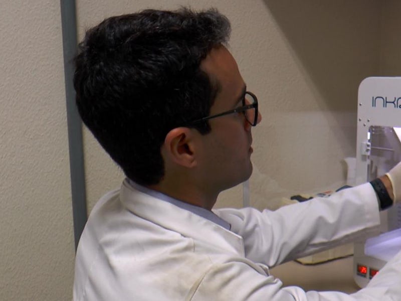
3D-printing human organs will save lives once perfected. Over 100,000 people are currently on a transplant waiting list, and 18 people die every day in the United States waiting to receive a transplant. If we could just print someone an organ using their own cells, they wouldn’t have to wait for a donor and there’d be pretty much no chance of the organ being rejected. Researchers in Brazil just successfully 3D-printed ‘mini-livers’ in a lab, and they function just like a regular liver. Their research was published in late November 2019 in the journal Biofabrication.
The researchers took blood from three volunteers and reprogrammed the blood cells to turn them into pluripotent stem cells, which are able to develop into any type of cell found in the human body.
The pluripotent stem cells were then induced to become liver cells, mixed with bioink and put through the 3D printer as spheroids. Spheroids are clusters of cells, so unlike other organ tissue that has been printed by researchers in the past, they were printing with more than one cell at a time. The researchers then let the structure culture for 18 days.
When the scientists examined the culture after it growth period, they found hepatic organoids, which are essentially miniature livers. They are able to function exactly as a liver does on a smaller scale.
Ernesto Goulart, a postdoctoral fellow at the University of São Paulo’s Institute of Biosciences and an author of the study, claimed in a statement that their method was more successful than other methods.
“Instead of printing individualized cells, we developed a method of grouping them before printing. These ‘clumps’ of cells, or spheroids, are what constitute the tissue and maintain its functionality much longer,” Goulart.
The tissue that was printed maintained hepatic functions longer than other liver tissue produced by 3D printing. Hepatic function is the term for how well the liver is working.
See also: How 3D-Printed Organs at the International Space Station May Cure Diseases
The researchers claim this whole process can be completed in 90 days. They claim their methods could definitely be used to print a fully functioning human liver.
“We did it on a small scale, but with investment and interest, it can easily be scaled up,” Goulart said.
Scientists have been getting much better at 3D-printing organ tissue and miniature versions of human organs in recent years. Scientists at Tel Aviv University were actually able to print a mini-human heart earlier this year. The bio-printing company Organovo is actually trying to have a patient receive a partial organ transplant with tissue made by a 3D printer by next year. A partial transplant would mean a portion of the organ tissue is replaced, which would in turn theoretically buy a patient a year or two before they need a transplant.
We’re still years away from a scenario where people are regularly getting entirely new organs that were made using their own cells, but it’s not science fiction anymore. Once this technology proliferates and becomes affordable for the average person, we’ll enter a time when people don’t die waiting for transplants anymore.
Abstract:
The liver is responsible for many metabolic, endocrine and exocrine functions. Approximately 2 million deaths per year are associated with liver failure. Modern 3D bioprinting technologies allied with autologous induced pluripotent stem cells (iPS)-derived grafts could represent a relevant tissue engineering approach to treat end stage liver disease patients. However, protocols that accurately recapitulates liver’s epithelial parenchyma through bioprinting are still underdeveloped. Here we evaluated the impacts of using single cell dispersion (i.e. obtained from conventional bidimensional differentiation) of iPS-derived parenchymal (i.e. hepatocyte-like cells) versus using iPS-derived hepatocyte-like cells spheroids (i.e. three-dimensional cell culture), both in combination with non-parenchymal cells (e.g. mesenchymal and endothelial cells), into final liver tissue functionality. Single cell constructs showed reduced cell survival and hepatic function and unbalanced protein/amino acid metabolism when compared to spheroid printed constructs after 18 days in culture. In addition, single cell printed constructs revealed epithelial-mesenchymal transition, resulting in rapid loss of hepatocyte phenotype. These results indicates the advantage of using spheroid-based bioprinting, contributing to improve current liver bioprinting technology towards future regenerative medicine applications and liver physiology and disease modeling.