Powerful X-Rays Appear to Reveal the Fossil Record's Most Ancient Bone
This is the mother of all bones.

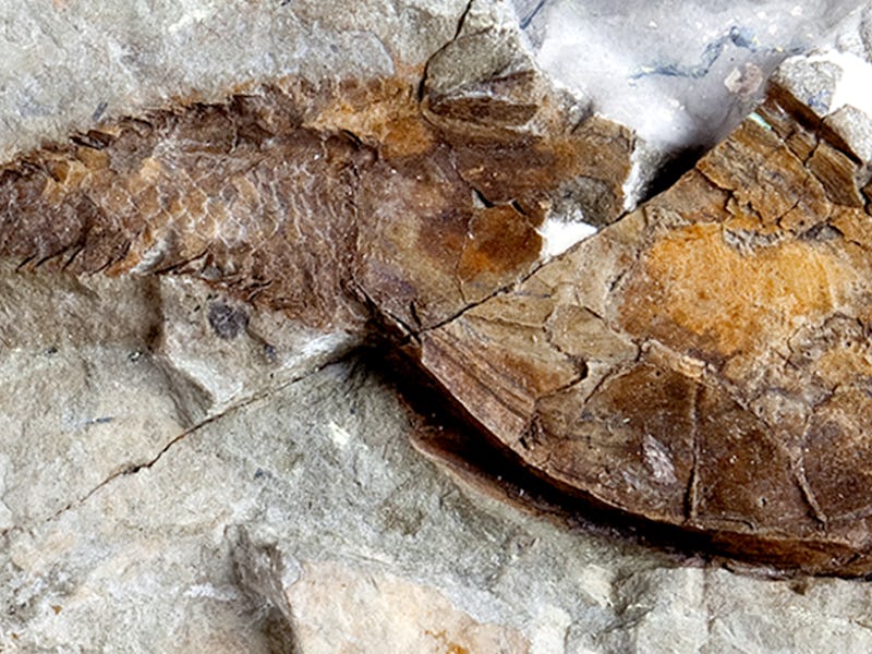
Over 400 million years ago, a strange, jawless fish swam in the world’s oceans. This fish had a flexible skeleton — a weird, bone-like material that wasn’t like present-day bone — that has defied categorization since its original owner died millions of years ago. On Tuesday, a study in Nature Ecology and Evolution reports that we’ve finally figured out what it is. It’s the most ancient example of bone in the entire fossil record.
The skeletal material seen in this ancient fish — part of a group called heterostracans — is called aspidin, the authors conclude. This material, explains study author Joseph Keating, Ph.D., a paleobiologist at the University of Manchester, has been nearly impossible to characterize because it doesn’t resemble any of the four tissue types — bone, cartilage, dentine, and enamel — that make up present-day bones and teeth. When biologists previously examined the aspidin fossils under a microscope, they were perplexed to find a crisscrossed branching structure.
The types of bone we know today don’t criss-cross under a microscope, so it’s been hard to figure out whether aspidin was actually bone. “For 160 years, scientists have wondered if aspidin is a transitional stage in the evolution of mineralized tissues,” says Keating. But his team’s detailed x-rays of the heterostracan fossils showed evidence that they likely represented a very important stage of bone evolution: the very first one.
A 2D image of an aspidin skeleton, note the weird branching pattern.
A major component of bone is an “organic matrix” of proteins like collagen, which come together to form a scaffold that minerals can attach to, turning the otherwise spongy tissue hard. Crucially, in the bones we’re used to, this matrix is usually structured in tubes that are linear, which is thought to be necessary in order for bone to mineralize.
Because of aspidin’s seemingly criss-crossed structure, researchers previously concluded that it couldn’t have those mineral components of the matrix. In other words, although it looked a lot like bone, it was probably not — likely just the evolutionary predecessor of mineralized bone.
Keating, though, decided to take an even closer look at aspidin. He spent over 100 hours scanning the fossil remains of heterostracan skeletons, using a technique called synchrotron tomography, which uses a type of X-ray so powerful it requires a particle accelerator to work. Keating found his particle accelerator at the Paul Scherrer Institute in Switzerland, where he used these high quality xrays to construct a three-dimensional model of the these aspidin skeletons.
Looking more closely than ever before, Keating found that the criss-crossing that had been so confusing in the past had disappeared. “I found that these tubes were strictly linear, lacking any kind of branching,” he wrote in a blog post in Nature. “The images from previous studies seem to be a result of 2-dimensional sectioning through tangled and overlapping tubes, giving the appearance of branching.”
This is what aspidin looks like in 3D
The 3D model revealed that the tubes actually were linear but appeared stacked on top of one another in random crisscrossed directions. For decades, he realized, as researchers looked at the tubes on two-dimensional X-rays, they appeared to be flattened out, forming a branching pattern that wasn’t indicative of their true structure. Crucially, the authors point out, these tubes house collagen, the scaffold protein that contributes to mineralization.
“We show that the spaces exhibit a linear morphology,” the authors write. “Instead, these spaces represent intrinsic collagen fibre bundles that form a scaffold about which mineral was deposited. Aspidin is thus acellular dermal bone.”
This tiny differentiation has staggering consequences when it comes to figuring out when mineralized skeletons, like the ones seen in humans, first evolved. By simply showing that these fish had mineralized skeletons, this team has set that date back a few million years:
“These findings change our view on the evolution of the skeleton,” concludes Phil Donoghue, Ph.D., co-author and paleobiologist from the University of Bristol. “We show that it is, in fact, a type of bone, and that all these tissues must have evolved millions of years earlier.”