Science and art are so often presented as diametrically opposed opposites, but, of course, there’s lots of overlap. Science is beautiful — not just in vague terms about the grand pursuit of knowledge, but actually aesthetically stunning. For the past 20 years, the Wellcome Image Awards have tried to honor and highlight “informative, striking and technically excellent images that communicate significant aspects of healthcare and biomedical science.”
The 22 winners for 2017 showcase the beautiful intricacies of science across several different fields of study, including biology, medicine, neuroscience, and beyond. Some are colorful, some are eerie, and all of them were created in an attempt to expand human understanding of the world we live in.
Scroll down to see the works of art and all of the ways that science and creativity can intertwine. You can also vote for your favorite for a chance to win a print of one of the stunning pieces.
Peter M Maloca, OCTlab at the University of Basel and Moorfields Eye Hospital, London; Christian Schwaller; Ruslan Hlushchuk, University of Bern; Sébastien Barré
Vessels of a healthy mini-pig eye
This model of a 3-D printed pig’s eye includes every tiny vessel that supplies blood to the eye — the smallest are about the diameter of a human hair. It is made out of the same material as Legos and took 39 hours to print.
Stephanie J Forkel and Ahmad Beyh, Natbrainlab, King’s College London; Alfonso de Lara Rubio, King’s College London
Language pathways of the brain
Also 3-D printed, this little bundle of axons mimics the pathway in the human brain that connects speech to language, called the arcuate fasciculus. It’s like the main telephone line in the brain and without it, we wouldn’t be capable of one of the most fundamental aspects of being human: verbal communication.
Gabriel Luna, Neuroscience Research Institute, University of California, Santa Barbara
Surface of a mouse retina
This veiny mosaic is an in-depth look at the retinal structure of a mouse’s eye. It was pieced together using more than 400 images of different optical scans, creating a complete map of the entire surface of the retina.
Suchita Nadkarni, William Harvey Research Institute, Queen Mary University of London
The Placenta Rainbow
In this color wheel, you’re actually looking at optical slices of pregnant mouse placentas. It might sound gross, but this image shows off the beautiful side of the nutrient-filled sack that feeds mammals before they pop out into the world. Scientists are using them to find solutions for pregnancy complications.
Ezequiel Miron, University of Oxford
Unravelled DNA in a human lung cell
Living creatures have flaws, even on a microscopic level. This gemstone-like beauty captures a digital image of two cells caught in a replication process called mitosis. For an unknown reason, they are stuck together, and the nucleus is getting pulled causing the DNA fibers to swirl around uncontrollably inside of it.
Gabriel Galea, University College London
Developing spinal cord
At the end of the day, we’re all just three little neural tubes. These tubes form in the first month of pregnancy and are the foundation for all human tissue. In this image, the tube to the left will form the spinal cord, brain, and nerves. The middle tube will come to form the skin, teeth, and hair. And the right tube will form the organs.
Ingrid Lekk and Steve Wilson, University College London
Zebrafish eye and neuromasts
This mutant zebrafish embryo is the product of CRISPR DNA editing. A foreign gene was inserted into the specimen, which expresses itself via glowing red fluorescence. It was designed to help scientists map instinctual behaviors like schooling.
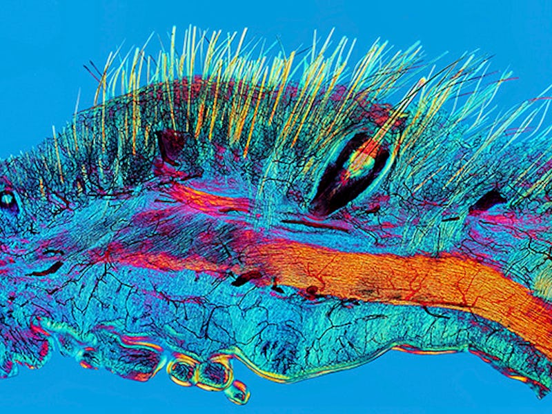
David Linstead
Cat skin and blood supply
Just further (funky) proof that our feline pets are natural-born killers. In this microscopic sliver of cat skin, the hairs and whiskers are indicated in yellow, the blood vessels are in black, and the red represents capillaries. Samples like this are leading scientists to believe that whiskers help cats feel vibrations in the air, signifying a moving object.
Mark Bartley, Cambridge University Hospitals NHS Foundation Trust
Intraocular lens ‘iris clip’
This image shows the wonders of a small clip inserted into the iris of an eye, — a common practice for helping people with nearsightedness and cataracts.
Joshua Mcdonald
Two young boys in rural Nicaragua
This photo documents two brothers on their way home from cutting in sugarcane fields in Nicaragua. They’ve lost their two older brothers to kidney failure, a common disease among men who work in hot temperatures.
Susan Smart
Patient receiving treatment during outreach eye screening in India
Here we see a patient being treated at a Unite for Sight clinic in India — a charity that has helped 1.9 million people worldwide, especially those in extreme poverty.
Eric Clarke, Richard Arnett and Jane Burns, Royal College of Surgeons in Ireland
#breastcancer Twitter connections
An interesting take on tweets about #breastcancer, this infographic uses cancerous-looking lumps to represent the connections between Twitter users who engaged in conversations about breast cancer. The large spot at the top represents two accounts that were mentioned thousands of times.
João Conde, Nuria Oliva and Natalie Artzi, Massachusetts Institute of Technology (MIT)
MicroRNA scaffold cancer therapy
These glowing strands are two bits of microRNA woven into a type of net. Scientists are hoping to use the net to trap cancerous tumors and deliver the MicroRNAs directly to them to stop them from growing.
Michael Northrop
Synthetic DNA channel transporting cargo across membranes
This biodesign illustration represents a DNA channel that acts as a two-way communication stream between a cell and it’s environment. The outer rings are the six helices and the blue orbs are the proteins that travel through them.
Madeleine Kuijper, Madeleine Kuijper Illustraties
Caricatural medieval medical practitioners
This digital illustration is inspired by medieval artwork, telling a story from the top left around clockwise to the bottom left. The story is about a man receiving very strange medical treatments. The snakes between the frame represent the ancient greek symbol for medicine, which is also seen today in the medical cross worn by doctors and first responders.
Original painting by Sophie McKay Knight, with imagery contributed by women scientists from the University of St Andrews – part of the Chrysalis project coordinated by Mhairi Stewart
‘Hidden Learning’, from the Chrysalis project
In this image, a girl wears a veil of sugar molecules on her head. As part of the Chrysalis project, which aims to bring women working in science together, the portrait represents women in science and the constant struggle they face when balancing work and home.
Spooky Pooka
Stickman – The Vicissitudes of Crohn’s (Resolution)
This stickman illustration is a visual representation of Chron’s Disease, a chronic illness that inflames the digestive system and often leads to painful symptoms, as represented by the sticks protruding from the figure’s abdomen.
Daria Kirpach
Rita Levi-Montalcini
This digital illustration depicts Rita Levi-Montalcini and her secret scientific endeavors during WWII in Italy. As a non-Aryan woman, she was forced to work in her home and it wasn’t until after the war that she was able to present her discovery of the nerve growth factor, which lead to our current understanding of tumors, deformations, and even dementia.
Mark R Smith, Macroscopic Solutions
Hawaiian bobtail squid
This close-up photograph of a baby bobtail squid draws the eye to the glowing black organ on its underside. The organ is actually an ink-filled sack, assisted by luminescent bacteria, which help the squid camouflage itself day or night.
Scott Birch and Scott Echols
Blood vessels of the African grey parrot
In this 3-D reconstruction of an African Grey Parrot, scientists can see the bird’s vascular system in great detail, including blood vessels in the head, neck, and beak.
Scott Echols, Scarlet Imaging and the Grey Parrot Anatomy Project
Pigeon thermoregulation
This image, which was also taken using the same technology as the previous picture, shows a pigeon’s vascular system, which sits just below the skin and helps the bird regulate its body temperature.
Collin Edington and Iris Lee, Koch Institute at MIT
Brain-on-a-chip
Stem cell research is a wondrous thing. In this microscopic image, the stem cells (pink) are sprouting nerve fibers (green). This kind of work is helping scientists understand how the environment affects the cell’s organization.